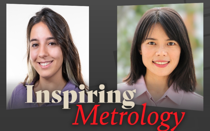
Inspiring Metrology: A conversation with Lei Zheng
Inspiring Metrology: A conversation with Lei Zheng, Researcher at Leibniz University Hannover
At this year’s EMO tradeshow in Hannover, we had the pleasure of meeting Lei Zheng, a distinguished researcher from Leipzig University. In a brief yet engaging interview, Lei gave us a sneak peek into her intriguing research project, sparking curiosity about the groundbreaking work in micro-nano production of optical elements.
During the interview, Lei highlighted the critical role of microscopy in her research. She mentioned how techniques like confocal microscopy and interferometry play a pivotal part in characterizing their samples, including steel structures, optical waveguides, and microresonators.
The S neox microscope, allows us to identify tiny features quickly and without damaging our samples, using a non-destructive approach.
Lei’s positive experience with the S neox system was evident as she pointed out its user-friendly software interface, swift measurement capabilities, and high-resolution image generation. Her practical insights as a two-year user of the system were valuable.
We invite you to watch the video to learn more about the cutting-edge advancements she and her team are making in the world of micro-nano production of optical elements.
Can you provide an overview of your current research project?
I’m working on a project called Excellent Cluster PhoenixD. In this big project, we have a lot of small projects. My task is mainly focused on the micro-nano production of optical elements. In optics and photonics, we need high-quality, high-resolution optical devices for different applications, for example, sensing and biophotonics.
How has microscopy contributed to the advancement of your research?
I would say it’s very useful and very helpful. In most cases, I use a confocal microscope and interferometry for our characterization. For example, we produce DOE (diffractive optical elements) structures and can easily get a 3D profile of this structure, which is very important for us.
Can you share any challenges that you have overcome thanks to optical microscopy?
Now we have a microscope with which we can quickly and easily characterize our samples. This is a really powerful tool. We can characterize our samples and generate high-quality and high-resolution images.
What is your favorite feature on the S neox?
This system has a lot of advantages, I would say.
The first one is like the software interface, so it’s really user-friendly, and everything is easy for the whole measurement process.
The next is the measurement process. It can be done in seconds. This is quick. And the last point is, again, the high-resolution measurement, . For example, a micro-resonator with a bus waveguide, which is a sample we produced for our project partners. If we want to achieve some optical performance, a small gap between the waveguide and the resonator is needed. We can produce samples with a few hundred nanometers gap size. With other microscope, it’s impossible to distinguish this gap. But with our system, we can see the gap.
The advantage of this point is this is a non-destructive approach, so we don’t need to destroy our sample.
What is your favorite technology, interferometry, confocal or focus variation?
I like them because they are all useful for our projects, and we use all the modules very often.
Could you provide some advice for other researchers who are interested in incorporating microscopy in their research?
First, I would say the whole equipment should be user-friendly and easy to handle.
Next is the equipment should support a quick measurement because this can help to save a lot of time.
The last is the equipment, or the microscope, should be able to produce high-quality and high-resolution measurements, especially for micro- and nanoproduction.


